Retinal Treatment
Overview
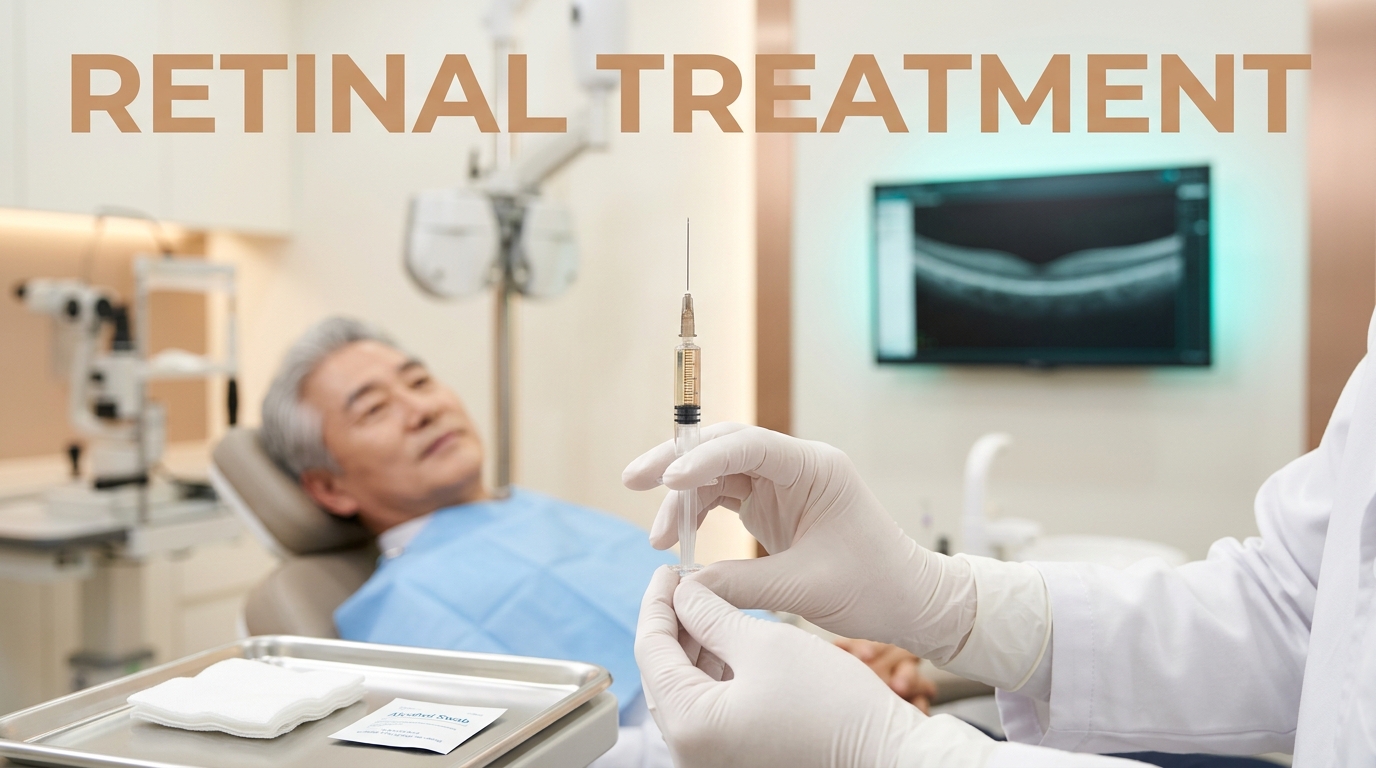
Retinal treatment refers to a group of specialized procedures aimed at managing disorders of the retina, the thin layer of tissue at the back of the eye that processes light. Common interventions include vitrectomy (removing the vitreous gel to access the retina), laser photocoagulation (sealing leaking vessels or tears), and scleral buckling (placing a band around the eye to support a detached retina).
These procedures are critical for preserving vision when the retina is compromised by trauma, aging, or systemic diseases like diabetes. Because the retina is neural tissue, these treatments focus on stabilizing the structure and preventing further vision loss rather than simply improving refractive errors like nearsightedness.
Expected Results
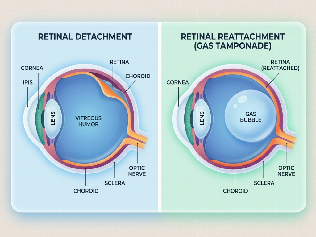
The primary goal of retinal treatment is the anatomical stabilization of the eye. For patients with retinal detachment, the objective is to reattach the tissue to its underlying blood supply. While many patients experience a stabilization or gradual improvement in vision, the final visual outcome depends heavily on whether the central part of the retina (the macula) was involved and how long the condition existed before surgery.
It is important to note that visual recovery is often a slow process that can take several months. In some cases, multiple procedures may be required to achieve the desired anatomical result, and some permanent vision loss may persist if the retinal cells were significantly damaged prior to intervention.
Why Korea & Cost
South Korea is well-regarded for its advanced ophthalmology centers, which utilize high-resolution imaging technologies like Optical Coherence Tomography (OCT) and ultra-widefield fundus photography. Korean retinal specialists often have extensive experience in micro-incisional vitrectomy surgery (MIVS), which uses exceptionally small instruments to minimize trauma and speed up healing.
The cost of retinal treatment in Korea is influenced by the complexity of the case, the specific surgical technique used (such as the use of silicone oil or gas tamponade), and the necessity for specialized inpatient monitoring. A comprehensive consultation and diagnostic imaging session are required to provide an accurate quote tailored to the patient's specific pathology.
Recovery & Aftercare
Recovery from retinal surgery is unique because it often requires strict head positioning. If a gas or air bubble is placed inside the eye to hold the retina in place, patients may need to maintain a face-down or side-lying position for several days to weeks. This is crucial for the success of the reattachment.
Patients will typically experience some redness, swelling, and a 'scratchy' sensation in the eye. Vision may be very blurry initially, especially if a gas bubble is present, as light cannot focus properly through the bubble. As the body naturally replaces the gas with fluid, vision gradually clears. Air travel is strictly prohibited until the gas bubble has completely dissipated.
Who Is It For?
- Individuals with sudden flashes of light or new floaters
- Patients diagnosed with retinal tears or detachment
- Individuals with advanced proliferative diabetic retinopathy
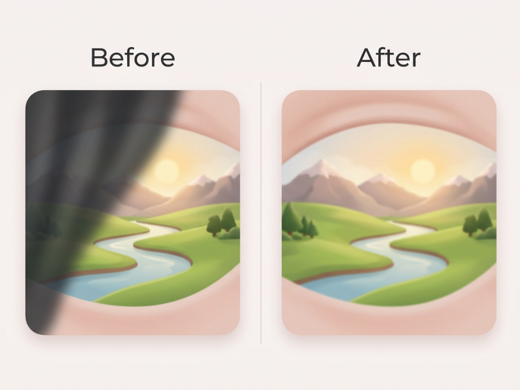
- Those experiencing a 'curtain' or 'shadow' over their field of vision
- Patients with macular holes or epiretinal membranes
Benefits
- High success rates for anatomical reattachment in many cases
- Prevention of permanent and total blindness
- Stabilization of vision in progressive retinal diseases
- Minimally invasive options available for certain tears
- Advanced diagnostic mapping for precise surgical targeting
How It's Done
- Administration of anesthesia and pupil dilation
- Creation of micro-incisions in the sclera (for vitrectomy)
- Removal of vitreous gel or repair of retinal tears using laser/cryotherapy
- Injection of air, gas, or silicone oil to support the retina
- Closure of incisions, often using self-sealing sutureless techniques
Risks & Side Effects
- Increased risk of cataract development post-surgery
- Potential for retinal redetachment requiring further surgery
- Temporary or permanent increase in intraocular pressure
- Endophthalmitis or internal eye infection
- Bleeding within the vitreous cavity
Recovery
- Strict face-down positioning may be required for 1-2 weeks
- Vision will be significantly blurred if gas or oil is used
- Redness and mild discomfort are common for the first 10 days
- Total recovery of vision can take 3 to 6 months
- Frequent follow-up visits are required to monitor eye pressure
Aftercare
- Administer prescribed antibiotic and anti-inflammatory eye drops
- Wear a protective eye shield while sleeping
- Avoid heavy lifting or strenuous exercise for at least one month
- Do not rub or apply pressure to the treated eye
- Monitor for sudden pain or a decrease in vision
Precautions
- Do not fly or travel to high altitudes if a gas bubble is present
- Avoid getting water or soap directly in the eye for the first week
- Ensure a caregiver is available to assist with positioning needs
- Inform the surgeon immediately of any sudden flashes or shadows
- Strictly adhere to the recommended head-down schedule
| Duration | 30-90 min |
|---|---|
| Recovery | 5-14 days |
| Anesthesia | Local anesthesia with sedation or General anesthesia |
| Price Range | Consultation required |





 Clinic Introduction
 Medical Facility
 Patient Care
 Consultation Room
 Medical Staff
 Recovery Suite
 Treatment Room